Compact Bone Diagram - Difference Between Compact and Spongy (with Table) - Ask .... Compact bone diagram bone cross section diagram file624 diagram of compact bone new. In long bones, as you move from the outer cortical compact bone to the inner medullary cavity, the bone transitions to spongy bone. As seen in the image below, compact bone forms the cortex, or hard outer shell of most bones in the body. A diagram of the anatomy of a bone, showing the compact bone. The cells of compact bone, which is also called cortical bone, appear to be tightly packed into a solid mass.
Compact bone is made of a matrix of hard mineral salts reinforced with tough collagen fibers. There are two types of bone tissue: It is also called osseous tissue or cortical bone and it provides structure and support for an organism as part of its skeleton, in addition to being a location for the storage of minerals like calcium.about 80% of the weight of the human skeleton comes from. Compact bone is formed in concentric circles. Anatomy of a long bone proximal epiphysis diaphysis distal epiphysis compact bone spongy bone medullary cavity.
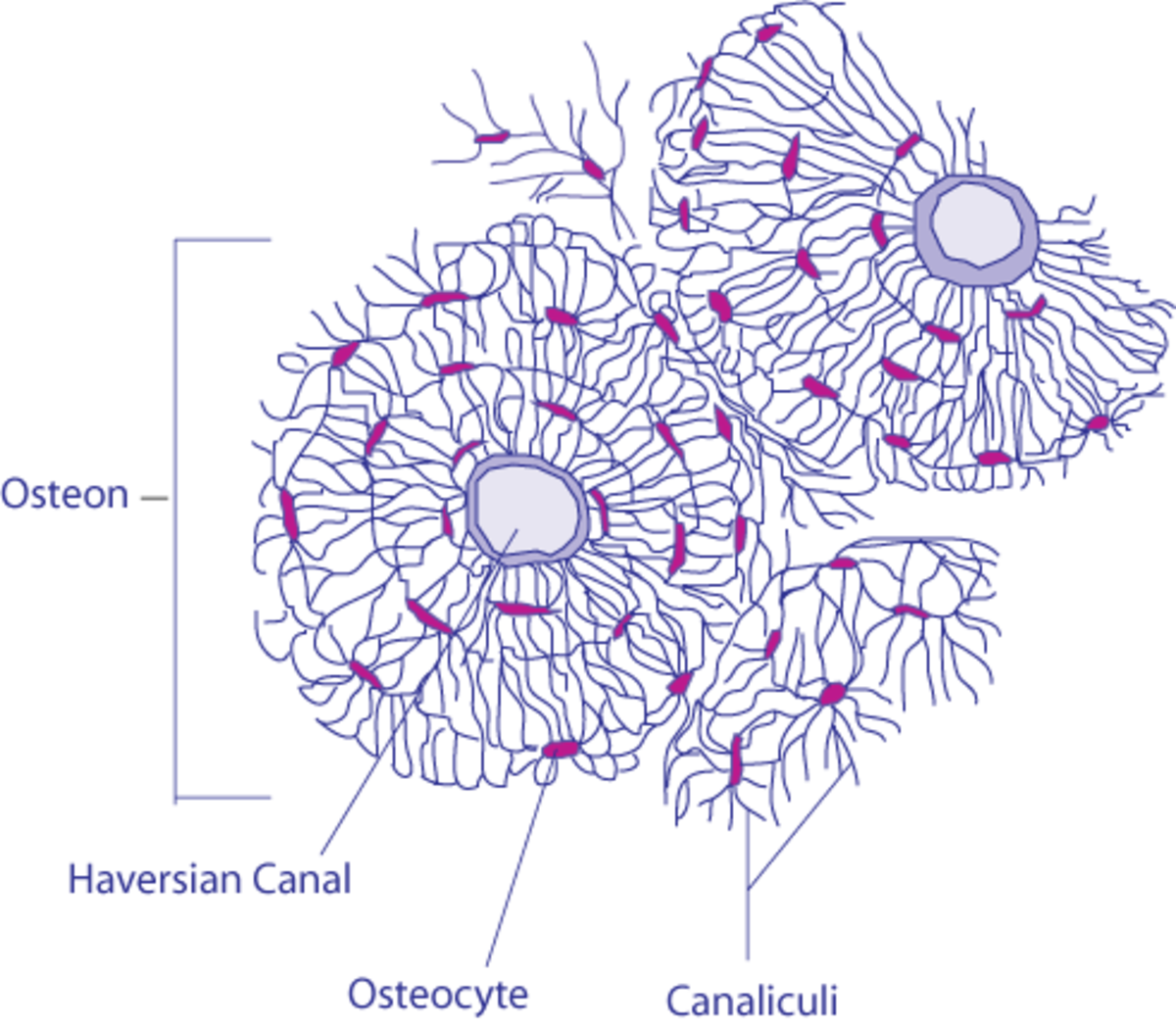
Osteoblasts, Osteoclasts, Calcium, and Bone Remodeling ... from usercontent2.hubstatic.com Compact bone is the denser, stronger of the two types of osseous tissue (figure 6.3.6). A diagram of the anatomy of a bone, showing the compact bone. Compact bone, also called cortical bone, dense bone in which the bony matrix is solidly filled with organic ground substance and inorganic salts, leaving only tiny spaces (lacunae) that contain the osteocytes, or bone cells.compact bone makes up 80 percent of the human skeleton; The main type of bone cell is the osteocyte (bone cell, shown as purple in the diagram). Separating the trabecular and compact bone is an epiphyseal plate (growth plate). Compact bone diagram osteon compact bone ap pinterest anatomy human anatomy and. Compact bones fills the outer layers of most of the bones. Compact bone, also called cortical bone, is the hard, stiff, smooth, thin, white bone tissue that surrounds all bones in the human body.
Spongy bones fills the inner layers of most of the bones.
The main type of bone cell is the osteocyte (bone cell, shown as purple in the diagram). Under magnification you can clearly see the system of concentric circles that forms compact bone. There are small canals that run through the bone, which allow blood vessels to penetrate it. Add to favorites 0 favs. As seen in the image below, compact bone forms the cortex, or hard outer shell of most bones in the body. Usually found in long bones of the body, it consists of units. The skeleton consists of bones and cartilages. (b) in this micrograph of the osteon, you can clearly see the concentric lamellae and central canals. Diagram of a typical long bone showing both cortical (compact) and cancellous (spongy) bone. The compact bones form the hard exterior of the bones, whereas the spongy bones have several pores that are filled with nerves and blood vessels. Compact bone is formed in concentric circles. The diagram above shows a longitudinal view of an osteon. Compact bone diagram bone cross section diagram file624 diagram of compact bone new.
Related posts of compact bone diagram labeled anatomical diagram of internal organs. In long bones, as you move from the outer cortical compact bone to the inner medullary cavity, the bone transitions to spongy bone. Compact bone is formed in concentric circles. Learn vocabulary, terms, and more with flashcards, games, and other study tools. Compact and spongy bone tissues share several similarities.
structures of a long bone to label - Google Search ... from i.pinimg.com Both are made of osteocytes, bone cells, and a mineral matrix that holds the osteocytes in. It is also called osseous tissue or cortical bone and it provides structure and support for an organism as part of its skeleton, in addition to being a location for the storage of minerals like calcium.about 80% of the weight of the human skeleton comes from. The remainder is cancellous bone, which has a spongelike appearance with numerous large spaces and is found in the. Although the calls are close together, this type of bone is not completely solid. (b) in this micrograph of the osteon, you can clearly see the concentric lamellae and central canals. You need to get 100% to score the 15 points available. The endosteum can be seen in the t.s. It makes up the outer cortex of all bones and is in immediate contact with the periosteum.
As seen in the image below, compact bone forms the cortex, or hard outer shell of most bones in the body.
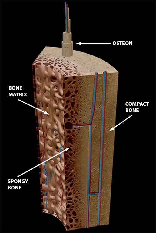
On the other hand, spongy bone is softer, and forms the inner layer of bones while covering a large surface area. The remainder is cancellous bone, which has a spongelike appearance with numerous large spaces and is found in the. Diagram of a typical long bone showing both cortical (compact) and cancellous (spongy) bone. Human bone generally comprises osseous tissue, an outer coating called a periosteum, and bone marrow. Compact bone is formed from a number of osteons, which are circular units of bone material and blood vessels. Compact bone, also called cortical bone, dense bone in which the bony matrix is solidly filled with organic ground substance and inorganic salts, leaving only tiny spaces (lacunae) that contain the osteocytes, or bone cells.compact bone makes up 80 percent of the human skeleton; Compact bone is made of a matrix of hard mineral salts reinforced with tough collagen fibers. Compact bone is the denser, stronger of the two types of osseous tissue (figure 6.3.6). Bone lamellae are arranged in regular haversian system. The diagram above shows a longitudinal view of an osteon. Although the calls are close together, this type of bone is not completely solid. Compact and spongy.the names imply that the two types differ in density, or how tightly the tissue is packed together. Usually found in long bones of the body, it consists of units.
Compact and spongy.the names imply that the two types differ in density, or how tightly the tissue is packed together. (b) in this micrograph of the osteon, you can clearly see the concentric lamellae and central canals. Compact bones fills the outer layers of most of the bones. Bone lamellae are arranged in regular haversian system. Many tiny cells called osteocytes live in small spaces in the matrix and help to maintain the strength and integrity of the compact bone.
In The Diagram Where Is The Osteon - Atkinsjewelry from www.visiblebody.com Online quiz to learn compact bone diagram; Under magnification you can clearly see the system of concentric circles that forms compact bone. Diagram of a typical long bone showing both cortical (compact) and cancellous (spongy) bone. Between the rings of matrix, the bone cells (osteocytes) are located in spaces called lacunae. The skeleton consists of bones and cartilages. A diagram of the anatomy of a bone, showing the compact bone. (b) in this micrograph of the osteon, you can clearly see the concentric lamellae and central canals. Compact and spongy.the names imply that the two types differ in density, or how tightly the tissue is packed together.
Diagram of a typical long bone showing both cortical (compact) and cancellous (spongy) bone.
Compact bone is the denser, stronger of the two types of osseous tissue (figure 6.3.6). Terms in this set (8) spongy bone (contains red marrow) compact bone (has osteons) osteon. (b) in this micrograph of the osteon, you can clearly see the concentric lamellae and central canals. Compact and spongy.the names imply that the two types differ in density, or how tightly the tissue is packed together. Illustration about compact bone, also called cortical bone, is the hard, stiff, smooth, thin, white bone tissue that surrounds all bones in the. Usually found in long bones of the body, it consists of units. There is also red bone marrow in the trabecular bone at both ends. Human bone generally comprises osseous tissue, an outer coating called a periosteum, and bone marrow. The main function of compact bone is to support the whole body, whereas spongy bones support the body structure. It is also called osseous tissue or cortical bone and it provides structure and support for an organism as part of its skeleton, in addition to being a location for the storage of minerals like calcium.about 80% of the weight of the human skeleton comes from. The remainder is cancellous bone, which has a spongelike appearance with numerous large spaces and is found in the. Compact bone is formed in concentric circles. Deep to the compact bone layer is a region of spongy bone where the bone tissue grows in thin columns called.